| Case report | Peer reviewed |
Cite as: Pollicino P, Gandini M, Perona G, et al. Use of Elastrator® rings to repair umbilical hernias in young swine. J Swine Health Prod. 2007;15(2):92–95.
Also available as a PDF.
SummaryThe objective of this study was to evaluate the efficacy and applicability of using Elastrator rings (Elastrator; Heiniger International, Switzerland) to repair umbilical hernias in swine. Ten 2- to 3-month-old gilts (Large White × Italian Landrace) with umbilical hernias were heavily sedated and placed in dorsal recumbency. After manual reduction of the hernia in each animal, two Elastrator rings were applied on the hernial sac, close to the abdominal wall. Animals showed no signs of post-treatment pain. In eight animals, the hernial sac separated at 21 to 28 days post treatment. These animals were marketed at a mean age of 220.5 ± 8.25 days and an average weight of 171 ± 7 kg. Repair was unsuccessful in two animals. The use of Elastrator rings to repair recently diagnosed umbilical hernias appears to be an effective method in commercial swine. The technique is simple and inexpensive, and animals need no special care. | ResumenEl objetivo de este estudio fue evaluar la eficacia y aplicabilidad del uso de anillos Elastrator (Elastrator; Heiniger International, Switzerland) para reparar hernias umbilicales en cerdos. Diez hembras de 2 a 3 meses de edad (Large White × Italian Landrace) con hernias umbilicales fueron fuertemente sedadas y colocadas en decúbito dorsal. Después de la reducción manual de la hernia en cada animal, se aplicaron dos anillos Elastrator en el saco hernial, cerca de la pared abdominal. Los animales no mostraron signos de dolor post tratamiento. En ocho animales, el saco hernial se separó a los 21 a 28 días post tratamiento. Estos animales fueron vendidos a una edad promedio de 220.5 ± 8.25 días y a un peso promedio de 171 ± 7 kg. La reparación fracasó en dos animales. El uso de los anillos de Elastrator para reparar hernias umbilicales recientemente diagnosticadas parece ser un método efectivo en cerdos comerciales. La técnica es simple y de bajo costo, y los animales no necesitan de cuidado especial. | ResuméL’objectif de la présente étude était d’évaluer l’efficacité et la faisabilité d’utiliser les anneaux Elastrator (Elastrator; Heiniger International, Suisse) pour la réparation d’hernies ombilicales chez le porc. Dix cochettes âgées de 2 à 3 mois (Large White × Landrace Italien) avec des hernies ombilicales ont été mises sous sédation profonde et placées en décubitus dorsal. Après réduction manuelle de l’hernie chez chaque animal, deux anneaux Elastrator ont été placés sur le sac herniaire, près de la paroi abdominale. Les animaux n’ont présenté aucun signe de douleur post-traitement. Chez huit animaux, le sac herniaire s’est détaché 21 à 28 jours post-traitement. Ces animaux ont été mis sur le marché à un âge moyen de 220.5 ± 8.25 jours et à un poids moyen de 171 ± 7 kg. La réparation n’a pas réussi chez deux des animaux. L’utilisation d’anneaux Elastrator afin de réparer les hernies ombilicales diagnostiquées tôt semble une méthode efficace chez les porcs commerciaux. La technique est simple et peu dispendieuse, et les animaux ne nécessitent aucun soin particulier. |
Keywords: swine, umbilical
hernia, Elastrator®
Search the AASV web site
for pages with similar keywords.
Received: March
14, 2006
Accepted: September
7, 2006
Umbilical hernia is one of the most common developmental defects in swine.1,2 Herniated pigs represent an economic loss, as they are generally sold at a lower price and slaughtered at a younger age due to the risk of ulceration and abscessation of the hernial sac or strangulation of the hernial contents.1,3,4 Hernia repair would permit recovery of affected animals and limit economic loss, although surgical intervention is expensive compared to the market value of the animal.3,4 As an alternative, Hall3 described a rapid and inexpensive method to reduce umbilical hernias in swine, consisting of topical application of concentrated nitric acid onto the hernial sac followed by isolation of the treated animal in a bedded pen for about 21 days. Another possibility for treatment of umbilical hernias is the application of Elastrators (Heiniger International, Switzerland) as described in foals.5 The aim of the present study was to assess whether the Elastrator technique could be used to reduce recently diagnosed umbilical hernias in young swine. This trial was authorized and conducted under the supervision of the Bioethical Committee of the Faculty of Veterinary Medicine of the University of Turin.
Case description
Ten Large White � Italian Landrace females with congenital hernias were purchased from a private farm at an average of 77.0 ± 13.5 days of age and average body weight of 49.1 ± 8.1 kg. These gilts would have been slaughtered at an early age on the farm of origin because of their hernias. They were housed in the training and experimental station of the Faculty of Veterinary Medicine, an indoor facility, at the University of Turin, and it was expected that after hernia repair, they would be slaughtered at the customary Italian market weight of 170 kg.
Animals were divided into two groups of five gilts, with each group housed in a 15-m2 pen with five other healthy females from the same source and of similar age and weight. Pens had atraumatic plastic-coated slotted metal floors and concrete walls. During the study, all animals were fed a commercial pelleted finishing ration. Relevant data for all animals are detailed in Table 1.
Table 1: Data collected 2 days before umbilical
hernia repair in 10 Large
* Maximal width and length measured by tape measure. † Diameter of internal hernia ring measured with a calliper. |
|||||||||||||||||||||||||||||||||||||||||||||||||||||||||||||||||||||||||||||||||||||||||||||||||||||
Two days before the trial began, the hernial sac of each study animal was examined for fistulas or skin lesions. At palpation, hernial sacs had a soft texture and the contents could be easily reduced. During this manipulation, animals showed no signs of pain or distress. Body temperatures were normal,6 ranging from 38.8�C to 39.1�C.
Application of Elastrators
Sedation was induced with azaperone, 4 mg per kg (Stressnil; Jannsen Pharmaceutical, Beerse, Belgium), administered by IM injection in the neck area. The five animals in each group were injected one after another. After sedation, each animal was placed on its right side and the width and length of the hernial sac were measured using a tape measure. Beginning 10 to 13 minutes after injection, each animal was placed in dorsal recumbency and the hernia was reduced by digital pressure. The diameter of the hernial ring was measured with a calliper. Subsequently, using a spring-loaded Elastrator applicator, two Elastrator rings were placed carefully around the hernial sac as close to the abdominal wall as possible. We found it helpful to grasp the hernial sac using atraumatic surgical forceps while the Elastrator rings were placed (Figures 1 and 2). The same procedure was applied to the second group of five animals. The mean time required to reduce hernias and place Elastrator rings was 2.25 ± 0.59 minutes per animal. At the end of the procedure, in order to allow treated animals to fully recover from sedation, all gilts were housed in individual pens for 12 hours before returning to their original pens.
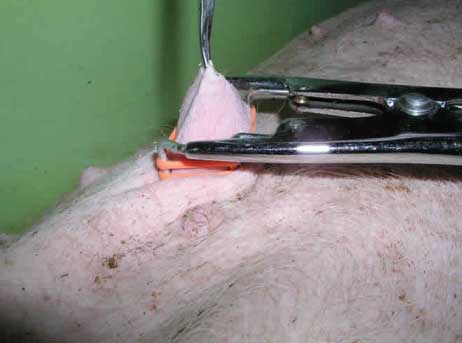
| Figure 1: Umbilical hernia repair using two Elastrator
rings. The gilt has been placed in dorsal recumbency after heavy sedation.
The hernia has been manually reduced, and the hernial sac is held up using
atraumatic forceps after placement of the Elastrator applicator.
|
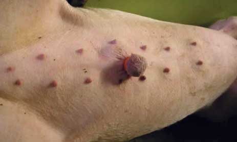
| Figure 2: Hernial sac after placement of Elastrator
rings.
|
Outcome of hernia repair using Elastrator rings
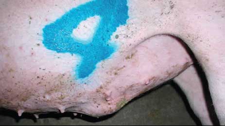
Animals were examined daily until the hernial sac separated from the abdomen. No behavioural differences were noted between treated animals and normal penmates. No signs of pain or discomfort or systemic signs were observed (eg, depression, inappetence, prolonged recumbency, self-isolation, self-mutilation, or cannibalism of hernial sacs) (Figure 3). Four to 5 days post treatment, the hernial sacs became noticeably discoloured (appeared cyanotic). Ischemic necrosis became evident 5 to 6 days post treatment and hernial sacs were completely necrotic by day 7 (Figure 4). During this period, animals showed no signs of pain or discomfort when hernial sacs were palpated. As observed in similar studies conducted in foals,5 transitory edema and swelling were observed around the area of Elastrator application between 5 and 21 days post treatment.
| Figure 3: Day 3 after placement of Elastrator rings
on an umbilical hernia.
|
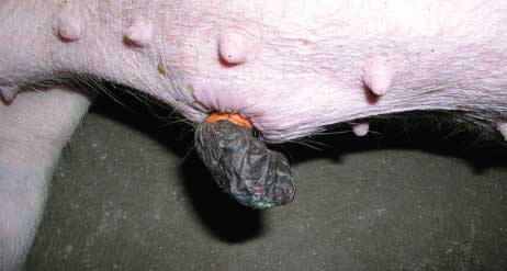
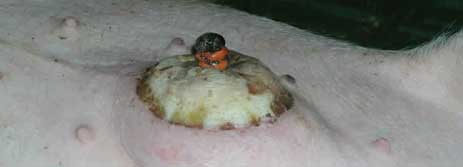
| Figure 4: Day 7 after placement of Elastrator rings
on an umbilical hernia, showing necrosis of hernial sac.
|
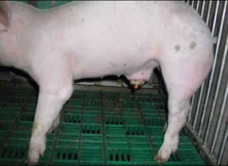
Recovery was uneventful for eight pigs. Separation of the hernial sac and the Elastrator occurred between 21 and 28 days post treatment (Figure 5). Average daily gain of these eight animals (791 ± 17 g) was comparable to that of the nonherniated penmates (802 ± 7 g). Mean slaughter weights of treated and healthy animals were 171 ± 7 kg and 177 ± 6 kg, respectively, at an average age of 220 ± 8 days.
| Figure 5: One of eight gilts successfully treated
for umbilical hernia using Elastrator rings. In each gilt, the necrotic
hernial sac separated from the abdomen at 21 to 28 days post treatment.
|
In two animals, treatment was unsuccessful. At day 7, edema developed between the Elastrator rings and the abdominal wall, pushing the Elastrator rings towards the distal tip of the hernial sac (Figures 6 and 7). These animals were sedated and underwent a second placement of Elastrator rings; however, hernias recurred a few days later in both animals.
| Figure 6: Gilt unsuccessfully treated for umbilical
hernia using Elastrator rings, showing the rings slipped to the distal
area of the hernial sac.
|
| Figure 7: Unsuccessful treatment of umbilical hernia
using Elastrator rings. Edema in the area where the rings were applied
caused them to slip toward the distal part of the hernial sac.
|
Discussion
These preliminary results indicate that reduction of recently diagnosed umbilical hernias with Elastrator rings may be an effective therapeutic method in commercial swine. A total of 2324 mg of azaperone (approximately $0.008 per mg; all currency in $US converted from Euro dollars) and 24 Elastrator rings (approximately $0.013 each) were used for 12 hernia repairs, an overall cost of approximately $18.90, with an average cost of approximately $2.36 for each successfully treated animal. The overall working time required to treat 10 animals was about 50 minutes, including the lag time between azaperone injection and hernia reduction and Elastrator application.
This technique appears cheaper, faster, and simpler to perform than other published methods.3,4 Moreover, in contrast to other methods,3,4 treated animals need no special care (eg, drug administration, individual housing during the healing period, bedded pens).
Further studies involving larger numbers of herniated pigs of both sexes and of different ages are in progress to better evaluate the feasibility of this technique under field conditions.
Implications
- Evidence from a small number of treated animals suggests that Elastrator rings may be useful for nonsurgical repair of umbilical hernias in commercial swine 2 to 3 months of age.
- Repair of umbilical hernias using a low-cost, nonsurgical method allows recovery of the economic value of successfully treated aniamls.
References
1. Searcy-Bernal R, Gardner IA, Hird DW. Effects of and factors associated with umbilical hernias in a swine herd. JAVMA. 1994;204:1660–1664.
2. Edwards MJ, Mulley RC. Genetic, developmental and neoplastic diseases. In: Straw BE, D’Allaire S, Mengeling WL, Taylor DJ, eds. Diseases of Swine. 8th ed. Ames, Iowa: Iowa State University Press; 1999:704–705.
3. Hall WH. Nonsurgical repair of umbilical hernias in swine. Mod Vet Pract. 1986;67:795–796.
4. St-Jean G, Anderson DE. Anesthesia and surgical procedures in swine. In: Straw BE, D’Allaire S, Mengeling WL, Taylor DJ, eds. Diseases of Swine. 8th ed. Ames, Iowa: Iowa State University Press; 1999:1139–1140.
5. Greenwood RES, Dugdale DJ. Treatment of umbilical hernias in foals with Elastrator rings. Equine Vet Educ. 1993;5:113–115.
6. Straw BE, Meuten DJ, Thacker BJ. Physical examination. In: Straw BE, D’Allaire S, Mengeling WL, Taylor DJ, eds. Diseases of Swine. 8th ed. Ames, Iowa: Iowa State University Press; 1999:5.